Assay Development

Assay Development

Transparency & constant communication are the principles that guide our assay development team and ensure your needs are met. Ultivue offers multiplex IF services through its proprietary InSituPlex® DNA-barcoding and antibody staining technology.


Explore our lab

Click on each station in the lab below to learn more about our assay development services.

Explore our lab

Click on each station in the lab below to learn more about our assay development services.

Assay Development

Our in-house team specializes in assay development, resulting in the rapid turnaround of custom assays and panels for any species. The development of non Ultivue menu markerscan come from commercially sourced, or customer provided reagents. With each project, image reviews and optimized assay conditions are provided to our customers.

Tissue preparation

At the start of any project, tissue undergo FFPE slide sectioning, for preparation ahead of staining. Our lab is overseen by trained histopathologists and histology scientists that can bring the necessary attention to detail required to support, develop, validate and implement your assay through the drug development pipeline.

Staining

Ultivue’s InSituPlex® mIF staining allows multiplexed staining of up to 12 biomarkers with H&E – on the same sample-using the Leica Bond Rx.

Our signal amplification technology creates a linear relationship of biomarker abundance and staining intensity with superior signal to noise ratio, ideal for image analysis.

Scanning & Imaging

Whole slide scanning & imaging are performed using the Zeiss Axioscan by trained technicians. As a CLIA-certified lab, we ensure regulatory compliance and data integrity for your projects. Rest assured that you will receive accurate, reliable, and timely results.

Analysis

Ultivue can provide complete phenotype analysis, advanced analysis and data mining based on your scientific question. With a complete representation of the tumor landscape including compartments, cell phenotypes and interactions, you areready for scientific exploration.

Final Report: Custom Data Delivery

At the end of each milestone, you will receive a report on the projects progress. Upon project completion, images, data results and final report are delivered by one of our data
sharing solutions.

Talk to our experts

Talk to our experts

Our pathologists, histopathology experts, and image analysis and AI scientists ensure that your scientific research needs are met.  The same experts are involved throughout the entire length of a project ensuring consistency from project inception all the way to delivery of the results.

Contact us
Your research goals are time-sensitive.

Let us support you in investigating your novel hypothesis by obtaining the most informative data from your valuable samples using our InSituPlex® technology and whole slide imaging (WSI). In partnership with our team of Histopathology experts, we will help advance your specific research needs.

H&E staining and mIF on one slide.

Instead of staining and assessing just one or two protein biomarkers at a time or even multiple markers throughout a set of serial sections, InSituPlex® technology allows for multiple Biomarkers coupled to traditional H&E staining to be visualized and quantified using just a single tissue slide.

Collaborative partnership for your assay development.

With In-house expertise in both IHC and multiplex immunofluorescence (mIF) assay development, customers can expect a unique experience with the Ultivue service team. Working in an agile and collaborative environment, get quick custom assay development and optimization.

Scientific expertise from start to finish.

The same expert IHC scientists are involved throughout the entire length of a project ensuring consistency from project inception all the way to delivery of the results.

CLIA-certified lab.

Ultivue is a CLIA-certified lab, ensuring regulatory compliance and data integrity for your projects. Rest assured that you will receive accurate, reliable, and timely test results.

Assay Development Milestones

01

Assay Development


· Antibody Verification
· Barcode conjugation
· Verification of conjugates

02

Panel Development

· Assay Development
· Optional sample testing

03

Staining & Imaging


· Verify sample integrity
· Stain samples
· Control testing

04

Image Analysis


· Phenotypes
· Regions
· Advanced analytics
· Data mining

Spatial phenomics expands the depth of information possible from a single tissue section

InSituPlex® Technology

Why Tissue Plex with InSituPlex®?

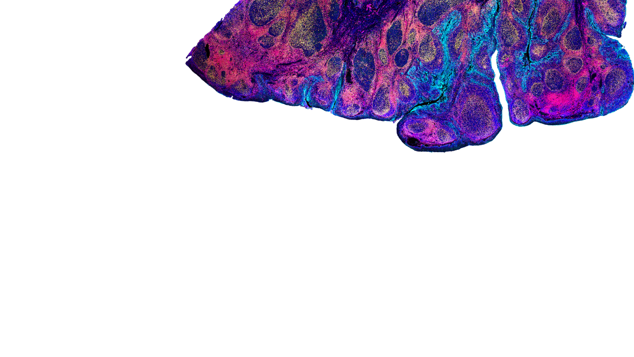
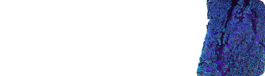
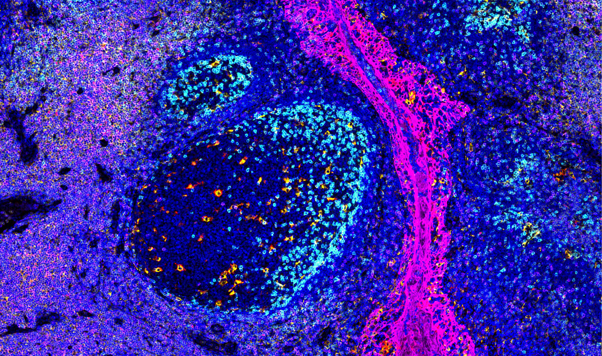
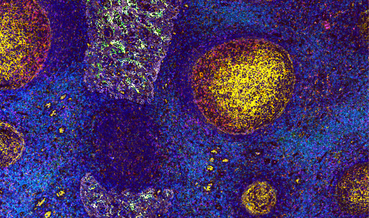
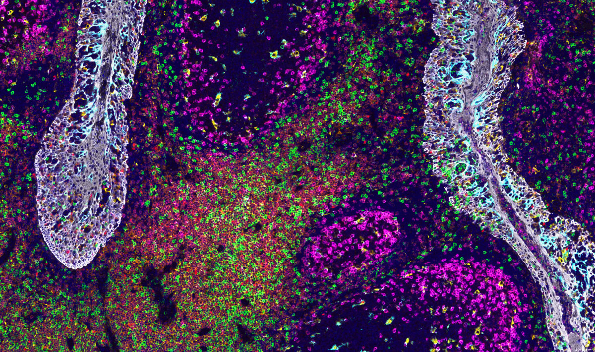
Ultivue provides researchers and scientists with multiplex biomarker assays and advanced image informatics for tissue phenotyping and digital pathology. Our proprietary InSituPlex® technology enables advanced exploration and interrogation of tissue samples for precision medicine research. These highly customizable solutions and scientific consultative approach strengthen and accelerate biomarker discovery and drug development programs.

InSituPlex® technology requires only a single antigen retrieval step before applying a mixture of DNA barcoded antibodies, which then bind to the desired array of antigens. After amplification, fluorescent probes are attached to those barcodes, allowing for visualisation of the model using fluorescent microscopy. The InSituPlex technology is available in 4, 8, 12-plex panel options.

Ultivue Staining Technology

Slide Preparation

The first step involves slide preparation (dewaxing) and single antigen retrieval from FFPE samples. This simplified process requires only one retrieval step, preserving the tissue.

Antibody Binding

The antibody barcode conjugates are incubated on the slide to stain the biomarkers. Each antibody is labelled with a DNA barcode that uniquely identifies it for detection. Up to 12 different antibodies can be added simultaneously. Only one single staining step is required, eliminating the need for secondary antibodies.

Barcode Amplification

During the next step the barcodes linked to the antibodies are amplified simultaneously and at the same rate. The extended barcodes do not interfere with the immediate surroundings so there is no steric hindrance or epitope masking.

Detection

The complementary fluorescent DNA probes are added and hybridize to the barcode targets. The samples are then ready for imaging.

Signal Removal

For additional rounds, signal must be removed by using exchange technology. This involves the gentle removal of fluorescent DNA probes, while keeping barcodes and tissue integrity intact. The slide is now ready for additional fluorescent DNA probes.

Not sure where to start?

We can help you to scope your project and discuss timelines and deliverables.

Assay Development | Image Analysis | Data Mining

Let’s speak with an expert
Not sure where to start?