


Immuno8 FixVUE™ Panel
If you‘re searching for a comprehensive view of the immune landscape, Immuno8 and ImmunoPro8 FixVUE® immuno oncology panels are the answer. With the use of 8 markers in one experiment, you can gain better insights into the tumor microenvironment.
Characterize T-cell subsets, immunosuppressive cells and interactions along the PD-L1/PD-1 checkpoint axis with this comprehensive immuno-oncology focused panel. The Immuno8 FixVUE™ panel enables phenotyping of at least 14 distinct cell types in the tumor microenvironment. Use this panel to dive deeper into the biology or find the key differences between samples and scale down to the essential markers to move your science forward.
View additional information (PDF)This antibody panel consists of the following markers:
Cell Phenotypes
Cell Phenotyping with the Immuno8 FixVUE™ Panel
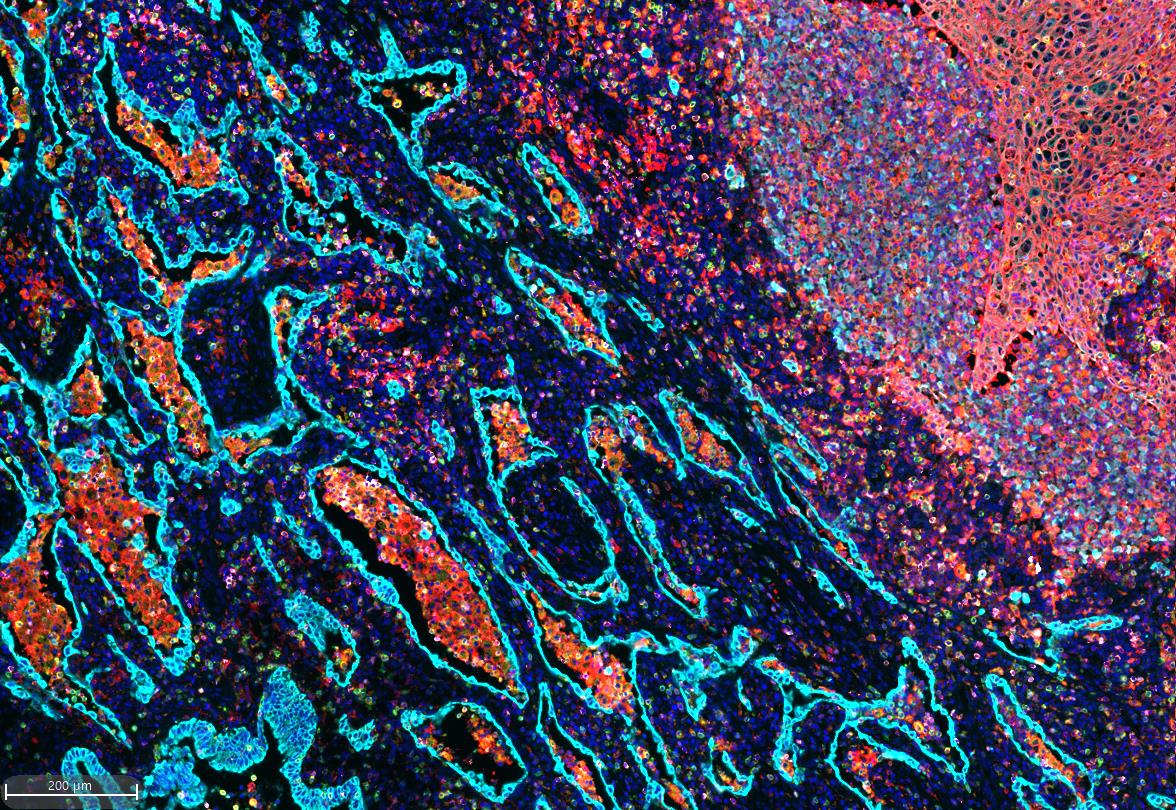
The Immuno8 FixVUE™ antibody panel enables users to identify and characterize T-cell subsets, immunosuppressive cells, and interactions along the PD-L1/PD-1 checkpoint axis. CD3 is a marker of T cells. CD4 is expressed on the surface of T helper cells. CD8 is a marker for cytotoxic immune cells (primarily cytotoxic T cells). CD68 is a marker of monocytes and macrophages. FoxP3 is a transcription factor and marker of cellular activation.
The co-expression of CD3, CD4, and FoxP3 indicates the regulatory T cell (T-reg) phenotype. PD-1 marks exhausted or suppressive T cells (CD3+/PD-1+) and can serve a pro-tumor function to help tumors evade the immune system. PD-L1 is an immune checkpoint marker that can be expressed on both macrophages (CD68+/PD-L1+) and tumor cells (CK/SOX10+/PD-L1). SOX10 is a marker of melanoma tumor cells while pan-cytokeratin detects carcinoma tumor cells.
Markers
Phenotype |
CD3 | CD4 | CD8 | CD68 | FoxP3 | PD-1 | PD-L1 | CK/SOX10 |
T cells |
|
|
|
|
|
|
|
|
T helper cells |
|
|
|
|
|
|
|
|
Cytotoxic T cell |
|
|
|
|
|
|
|
|
FoxP3+ T cells |
|
|
|
|
|
|
|
|
Exhausted T cells |
|
|
|
|
|
|
|
|
Regulatory T cells (T-reg) |
|
|
|
|
|
|
|
|
CD4/CD8 Double-positive T cells |
|
|
|
|
|
|
|
|
CD8+ Regulatory T cell (CD8 T-reg) |
|
|
|
|
|
|
|
|
Exhausted, Cytotoxic T cell |
|
|
|
|
|
|
|
|
Macrophage |
|
|
|
|
|
|
|
|
Immunosuppressive macrophage |
|
|
|
|
|
|
|
|
Tumor cell |
|
|
|
|
|
|
|
|
Immune-evading tumor cell |
|
|
|
|
|
|
|
|
This multiplexed immunofluorescence (mIF) panel allows for the spatial identification of single biomarkers and co-expression of multiple markers in cells enabling the observation of several biologically relevant phenotypes. The above is a partial of the 256 distinct binary phenotypes that this panel can identify. The number of phenotypes increases if binned marker intensities are taken into account (e.g.PD-1, PD-L1, FoxP3 expression levels)
Phenotype
Markers
Markers
Phenotype, Markers
T cells
T helper cells
Cytotoxic T cell
FoxP3+ T cells
Exhausted T cells
Regulatory T cells (T-reg)
CD4/CD8 Double-positive T cells
CD8+ Regulatory T cell (CD8 T-reg)
Exhausted, Cytotoxic T cell
Macrophage
Immunosuppressive macrophage
Tumor cell
Immune-evading tumor cell
Product Biology
Markers
Main Cell Type
Markers,Main Cell Type
CD3
T cells
CD3, T cells
Identifies all T cells and is the most specific marker for T cells, including lineage based markers such as CD4 and CD8.
CD4
Helper T cell
CD4, Helper T cell
CD4 binds to MHC class II molecules and participates in signal transduction processes. Its expression is used to identify helper T cells of which there are many different subsets each contributing to the overall immune function through their unique cytokine profile.
CD8
Cytotoxic T cells
CD8, Cytotoxic T cells
Cytotoxic T cells are responsible for mediating apoptosis of cancer cells through a variety of mechanisms that include the release of granzyme B from the T cells.
CD68
Marophages
CD68, Marophages
CD68 is expressed on human macrophages and other mononuclear phagocytes. CD68 functions in phagocytic activities and macrophage homing. An increased CD68+ macrophage index is associated with metastasis, poor prognosis, and reduced overall survival in multiple types of cancer.
FoxP3
Regulatory T cell
FoxP3, Regulatory T cell
FoxP3 is a transcription factor important in the development and inhibitory function of regulatory T cells (Tregs). FoxP3 functions by inhibiting cytokine production and T cell effector function, thus playing a crucial role in maintenance of immunological tolerance and control of immune responses against tumors and pathogens.
PD-1
Exhausted T cell
PD-1, Exhausted T cell
Programmed death receptor-1 (PD-1), an immune checkpoint molecule, is over expressed in cancer, leading to increased T cell exhaustion and a decreased anti-tumor response.
PD-L1
T cells, Monocytes, NK cells and Macrophages
PD-L1, T cells, Monocytes, NK cells and Macrophages
PD-L1 is expressed on the surface of tumor cells and it is able to bind to PD-1 on the surface of activated T cells and B cells. The binding of PD-L1 to PD-1 leads to an immunosuppressive effect and allows the tumor to evade immune destruction.
CK/SOX10
Tumor cells
CK/SOX10, Tumor cells
Cytokeratins are expressed in cells of an epithelial origin including most carcinomas. Sox10 is expressed in cells derived from the neural crest including melanocytes that give rise to melanomas.
|
|
