


PD-1 FixVUE™ Panel
Determine whether the immune response has exhausted. Our Immune exhaustion 4-plex/5-color panel enables co-localization of T cells, memory T cells, tumor cells, and the underlying PD-1 checkpoint immune exhaustion mechanism at play.
View additional information (PDF)This antibody panel consists of the following markers:
Cell Phenotypes
Cell Phenotyping with the PD-1 FixVUE Panel
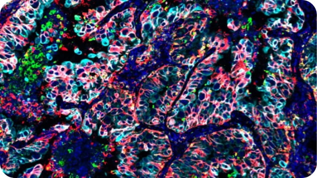
The PD-1 FixVUE antibody panel enables users to profile tumor samples by detecting the location and abundance of memory T cells and exhausted T cells. CD3 is a general marker for T cells. CD45RO is a marker for memory T cells, which have been primed against antigens and can provide anti-tumor immune responses. PD-1 marks exhausted or suppressive T cells and can serve a pro-tumor function to help tumors evade the immune system. SOX10 is a tumor marker for melanomas while PanCK detects carcinomas; antibodies for PanCK and SOX10 are provided as a cocktail in this panel.
Markers
Phenotype |
CD3 | CD45RO | PD-1 | PanCK/SOX10 |
T cells |
|
|
|
|
Memory cells |
|
|
|
|
Exhausted cells |
|
|
|
|
Carcinoma or Melanoma |
|
|
|
|
Memory T cells |
|
|
|
|
Exhausted memory cells |
|
|
|
|
Exhausted T cells |
|
|
|
|
Exhausted memory T cells |
|
|
|
|
Phenotype
Markers
Phenotype, Markers
T cells
Memory cells
Exhausted cells
Carcinoma or Melanoma
Memory T cells
Exhausted memory cells
Exhausted T cells
Exhausted memory T cells
Product Biology
Markers
Main Cell Type
Markers,Main Cell Type
CD3
T cells
CD3, T cells
Identifies all T cells and is the most specific marker for T cells, including lineage based markers such as CD4 and CD8.
CD45RO
Memory T cell
CD45RO, Memory T cell
Indicates that the T cell has previously encountered its cognate antigen. An increase in CD45RO+ T cells in tumors is associated with a better prognosis.
PD-1
Exhausted T cell
PD-1, Exhausted T cell
A marker of T-cell exhaustion indicating that normal function of the cell has been disrupted. May alsoindicate an immuno-suppressive phenotype. Increased levels of PD-1+ T cells are associated with a poorer prognosis. PD-1 is the target of multiple therapeutics.
panCK/ SOX10
Tumor cells
panCK/ SOX10, Tumor cells
The panel may be used to interrogate many tumor sample types for Research. A cocktail of optimized reagents for the detection of pan-Cytokeratin and Sox10 protein markers is provided. Cytokeratins are expressed in cells of an epithelial origin including most carcinomas. Sox10 is expressed in cells derived from the neural crest including melanocytes that give rise to melanomas.
