


PD-L1 FixVUE™ Panel
Determine whether the tumor is “hot” or “cold”.
Our Immune infiltration 4-plex/5-color panel enables co-localization of macrophages, cytotoxic T cells, tumor cells, and the underlying inhibitory or inflammatory mechanisms at play along the PD-L1 axis.
This antibody panel consists of the following markers:
Cell Phenotypes
Cell Phenotyping with the PD-L1 FixVUE™ Panel
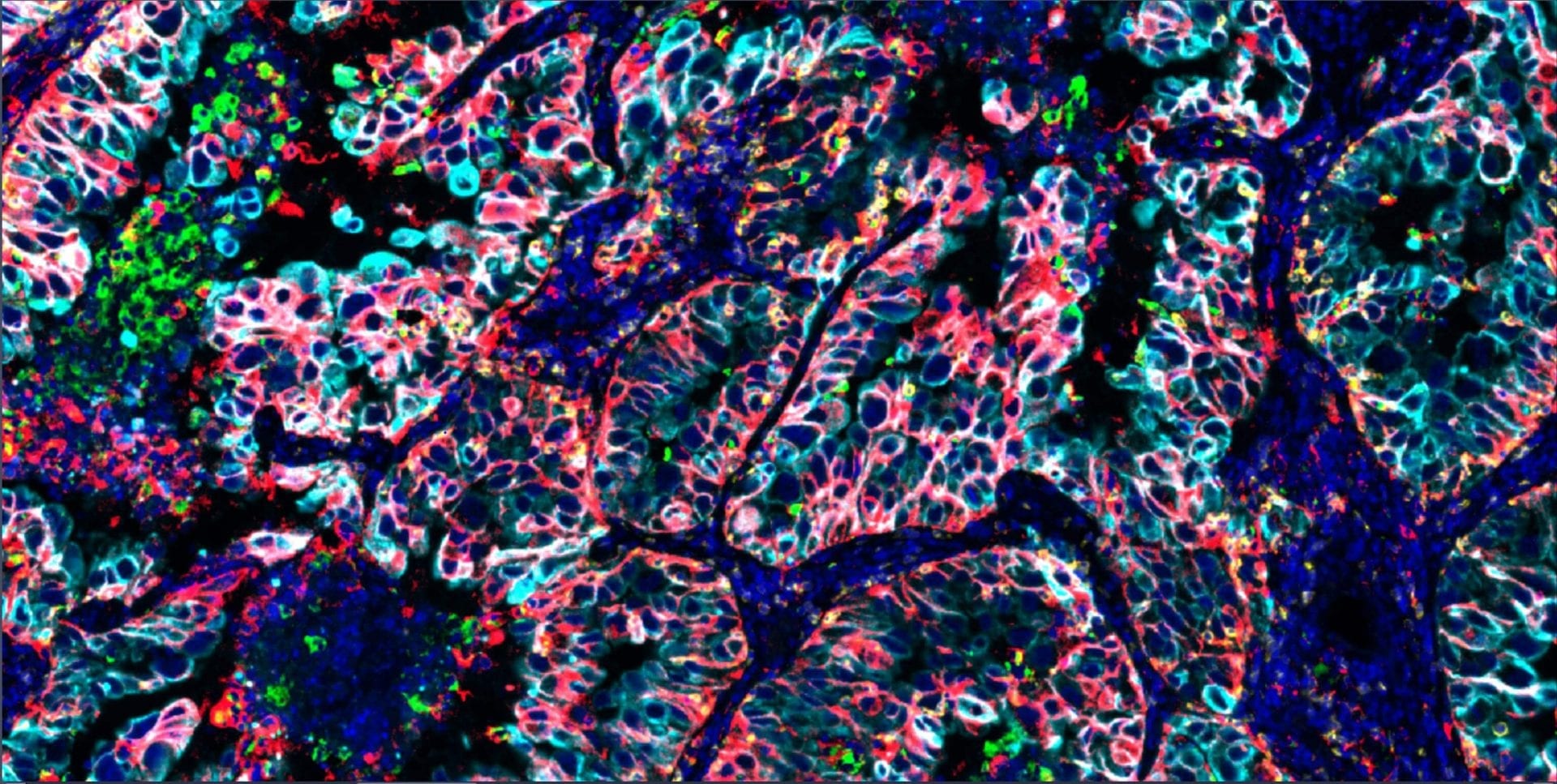
The antibody panel in the PD-L1 FixVUE enables users to detect immune infiltration and profile tumor samples. CD8 is a marker for cytotoxic immune cells (mainly cytotoxic T cells). CD68 is a marker for macrophages. PD-L1 is an immune checkpoint marker that can be expressed on both macrophages and tumor cells. SOX10 is a tumor marker for melanomas while panCK detects carcinomas; antibodies for panCK and SOX10 are provided as a cocktail in this panel. The expression level of PD-L1+ tumor cells and their proximity to CD8+ T cells have been highlighted as potential predictors of therapeutic response.
Markers
Phenotype |
CD8 | CD68 | PD-L1 | PanCK/SOX10 |
Cytotoxic immune cells |
|
|
|
|
Macrophages |
|
|
|
|
PD-L1 checkpoint expression |
|
|
|
|
Carcinoma (PanCK) or Melanoma (SOX10) |
|
|
|
|
Immunosuppressive macrophages |
|
|
|
|
Immune evading tumor cells |
|
|
|
|
Phenotype
Markers
Phenotype, Markers
Cytotoxic immune cells
Macrophages
PD-L1 checkpoint expression
Carcinoma (PanCK) or Melanoma (SOX10)
Immunosuppressive macrophages
Immune evading tumor cells
Product Biology
Markers
Main Cell Type
Markers,Main Cell Type
CD8
Cytotoxic T cells
CD8, Cytotoxic T cells
Cytotoxic T-cells are responsible for mediatingapoptosis of cancer cells through the release of perforin and granzyme B from the T-cells. Increased levels of PD-L1+ cells alone in tumor samples provide limited differentiation between samples. However, the proximity of PD-L1+ cells to CD8+ cells may support better differentiation.
CD68
Marophages
CD68, Marophages
Macrophages modulate the immune response.
PD-L1
Checkpoint protein
PD-L1, Checkpoint protein
Allows cells to escape immune surveillance by binding to PD-1. A search of ClinicalTrials.gov yields nearly 700 clinical trials with the keyword PD-L1. The association of PD-L1 expression with clinical outcome is uncertain at this time, but researchers are working to establish the relationship of this marker to other markers such as CD8.
PanCK/ SOX10
Tumor cells
PanCK/ SOX10, Tumor cells
A cocktail of optimized reagents for the detection of pan-Cytokeratin and Sox10 protein markers is provided. Cytokeratins are expressed in cells of an epithelial origin including most carcinomas. Sox10 is expressed in cells derived from the neural crest including melanocytes that give rise to melanomas.
