


Presented at: /
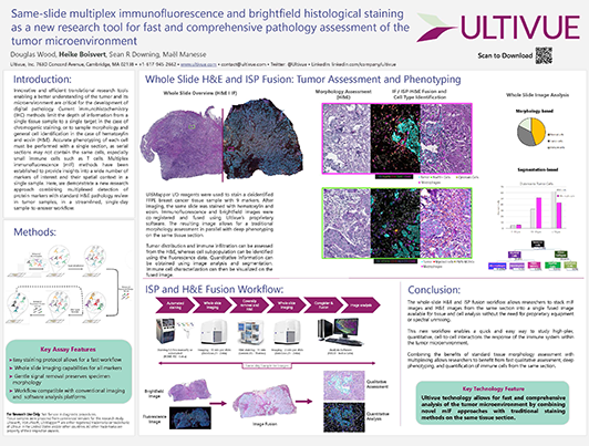
Same-slide multiplex immunofluorescence and H&E staining
Register here for download
Relevant for
FixVUE, InSituPlex
Description Innovative and efficient translational research tools enabling a better understanding of the tumor and its microenvironment are critical for the development of digital pathology. Current immunohistochemistry (IHC) methods limit the depth of information from a single tissue sample to a single target in the case of chromogenic staining, or to sample morphology and general cell identification in the case of hematoxylin and eosin (H&E). Accurate phenotyping of each cell must be performed with a single section, as serial sections may not contain the same cells, especially small immune cells such as T cells. Multiplex immunofluorescence (mIF) methods have been established to provide insights into a wide number of markers of interest and their spatial context in a single sample. Here, we demonstrate a new research approach combining multiplexed detection of protein markers with standard H&E pathology review in tumor samples, in a streamlined, single-day sample-to-answer workflow
Autors
Douglas Wood, Heike Boisvert, Sean R. Downing and Maël Manesse

Want to meet with us? Check out our upcoming events.
Related Content
Use of Ultivue InSituPlex®Multiplex Immunofluorescence to Localize and Quantify Regulatory T Lymphocytes in Crohn’s Disease and Ulcerative Colitis
A phase I clinical trial on intratumoral administration of autologous CD1c (BDCA-1)+ myeloid dendritic cells (myDC) plus talimogene laherparepvec (T-VEC) in patients with advanced melanoma
Overview of the differences between standard IHC and multiplexed immunofluoresence
Ultivue
Analysis of macrophages to the overall PD-L1 microenvironment using TMAs and InSituPlex
Presented at ASCO 2022

