


Jianghua Wu et. al., 2021
Validation of mIF and digital image analysis for PD-L1 expression and immune cell assessment
Relevant for
Biotech, Pharma, CROs, AMCs
Abstract. This study aimed to validate the application of combined multiplex immunofluorescence (mIF) and digital image analysis (DIA) in formalin-fixed and paraffin-embedded tissues for the quantitative assessment of programmed death-ligand 1(PD-L1) and immune cells (ICs) in non-small cell lung cancer (NSCLC).
Methods. Fifty resected samples of NSCLC were sequentially stained with a DNA-tagged mIF (panel including PD-L1, CKpan, CD8, CD68 and 4′,6-diamidino-2-phenylindole (DAPI)) and conventional immunohistochemistry (cIHC). The assessment of cell density and consistency of tumor proportion score (TPS) via DIA were compared with those by pathologists.
Results. A strong correlation in the cell population of immune markers was obtained between mIF and cIHC (for PD-L1: R=0.9304, CKpan: R=0.8231, CD8: R=0.9314 and CD68: R=0.8366) within 95% limits of agreement. The continuous TPS calculated using mIF was highly consistent with the IHC staining results which were evaluated by pathologists (R=0.9362). However, in the comparison of TPS using interval variables, a poor agreement was obtained at a cut-off of 1% (κ=0.197), whereas excellent agreement was achieved at cut-offs of 50% (κ=0.908) and 5% (κ=0.823). DIA on mIF showed that PD-L1 commonly colocalized with CD68+ macrophages and CD8+ cytotoxic cells were closer to PD-L1-/CK+ tumor cells (TCs) than to PD-L1+/CK+ TCs in spatial distribution.
Conclusions. A combination of mIF and DIA is useful for the quantification of PD-L1 expression and IC populations in NSCLC. Further validation of TPS at a cut-off of 1% and assay harmonization is essential for translating this method in a diagnostic setting.
What is the problem?
IHC is not sufficient enough to generate the necessary information from the comprehensive landscape of the TME
When we consider digital pathology applications most of our current knowledge has been gleaned from Immunohistochemistry (IHC). While this technique has helped identify targets and cell types as part of the tumor microenvironment (TME), the data has been limited by laborious protocols to only 1-2 markers. Its become clear that IHC is not sufficient enough to generate the necessary information from the comprehensive landscape of the TME and indeed to date, one has to superimpose multiple slides of data to achieve the level of multiplexing required.
Current clinical stratification for NSCLC immunotherapy regimens requires a positive measurement of tumor cell surface PD-L1 expression and a derived tumor proportion score (TPS) score. However co-localization analysis of the immune checkpoint marker PD-L1 and other immune markers ideally requires the detection of multiple markers on the same tissue slide. The goal of the study was to perform a comparison of DNA-barcoding mIF using Ultivue’s InSituPlex technology and conventional IHC to assess PD-L1 status and other immune cell types in NSCLC tumors
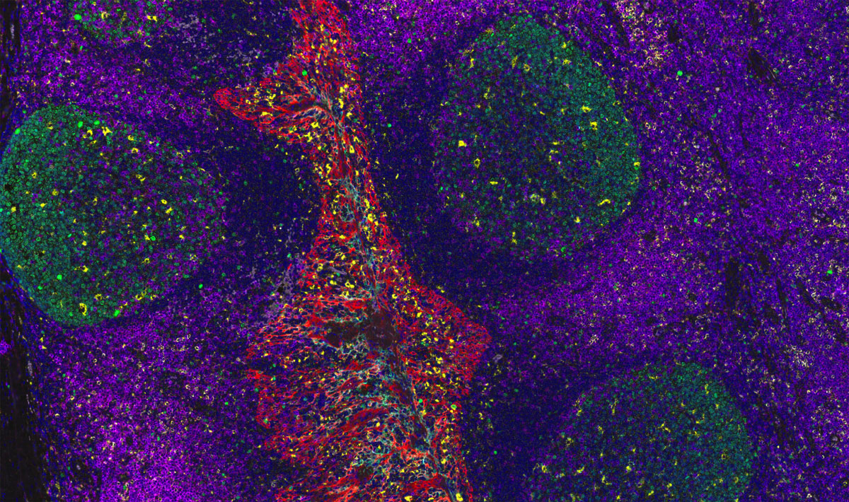
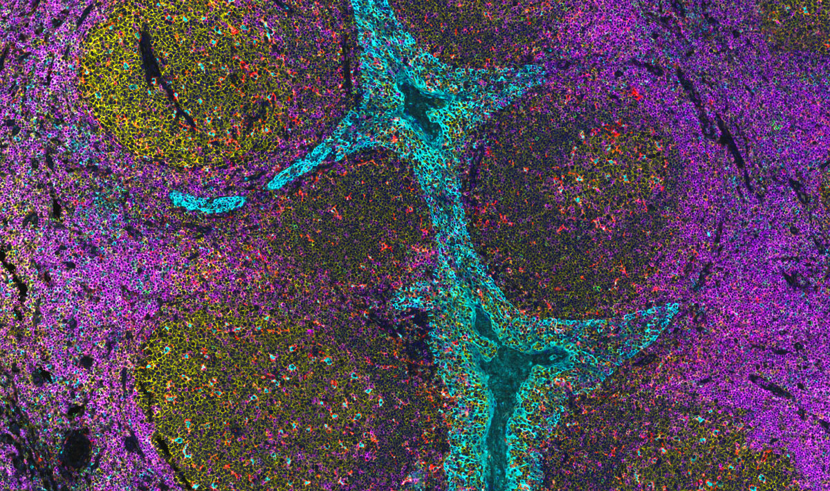
Multiplex immunofluorescence (mIF) and the use of digital image analysis is useful for the quantitative assessment of PD-L1 and other immune cells and is highly consistent with traditional single marker IHC staining results. However,the implementation of mIF and digital image analysis provides a unique insight into cell subsets, specific phenotype co-localization and spatial relations of immune checkpoints (like PD-L1) to tumor cells.
Results
Greater detail and insights into cellular identity and biomarker expression were achieved using mIF
Traditional IHC of PD-L1 was performed using the PD-L1 kit (PD-L1 22C3, Dako) and multiplexed PD-L1 together with expression of CD68, CD8 and PanCK/Sox10 was enabled by the Ultivue FixVUE PD-L1 pre-optimized kit.
Findings from the study demonstrated a high correlation in immune cell densities and TPS between IHC and mIF. However greater detail and insights into cellular identity and biomarker expression were achieved using mIF. Specifically, simultaneous detection of both PD-L1+ and cytokeratin epithelial cell marker+ (CK+) using ISP technology enabled differentiation of PD-L1+ tumor cells from PD-L1+ T cells and PD-L1+ macrophages.
Further, detection of PD-L1+CK+ and CK+ cells permitted calculation of a quantitative rather than a qualitative TPS. Studies also showed that the calculation of TPS using mIF compared favorably to the TPS calculated using IHC and pathologist review.
Start finding your answers. Talk to us to define your next project.
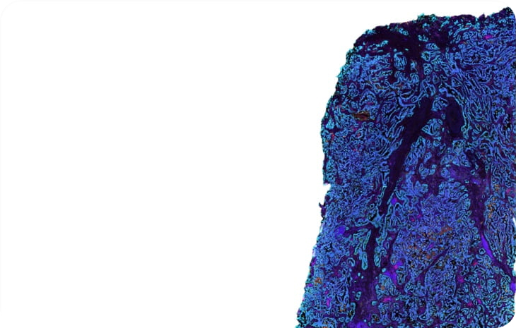
Images
Lorem ipsum dolor sit amet, consetetur sadipscing elitr, sed diam nonumy eirmod tempor invidunt
Lorem ipsum dolor sit amet, consetetur sadipscing elitr, sed diam nonumy eirmod tempor invidunt
Lorem ipsum dolor sit amet, consetetur sadipscing elitr, sed diam nonumy eirmod tempor invidunt
References
Wen J. et. al. A pan-cancer analysis revealing the role of TIGIT in tumor microenvironment. Sci Rep. 2021 Nov 18;11(1):22502.
