


Presented at: AACR 2022
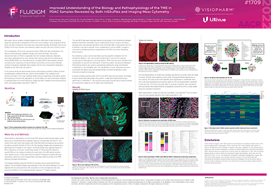
Improved Understanding of the Biology and Pathophysiology of the TME in PDAC Samples Revealed by Both InSituPlex and Imaging Mass Cytometry
Register here for download
You are currently viewing a placeholder content from HubSpot. To access the actual content, click the button below. Please note that doing so will share data with third-party providers.
More InformationRelevant for
Collaboration, FixVUE, Imaging mass cytometry, InSituPlex
Description This poster demonstrates a unique tissue phenotyping workflow combining three complementary methods that can unravel novel insights in the complex tumor microenvironment. The highlighted workflow delivers tissue morphology information, spatial phenotyping of immune cell population on whole slides, and high-dimensional imaging in selected regions of interest (ROIs) by combining H&E, multiplex immunofluorescence (mIF), and Imaging Mass Cytometry™(IMC™) together with bespoke data analysis.
Authors
Andrew Quong, Nina Lane, Derek Quong, Gourab Chatterjee, Devan Fleury, Kirsteen H. Maclean, Maël Manesse, Keith A. Wharton Jr, Fabian Schneider, Dan Winkowski and James Mansfield.

Want to meet with us? Check out our upcoming events.
Related Content
Spatial multi-omics analysis targeting protein and RNA biomarkers on a single FFPE tissue section using an integrated staining and imaging workflow
Presented at AACR 2022
Informing Spatial Biology with Multiplexed Immunofluorescence: A Pathologist’s Perspective
60 minutes
A workflow for cloud-based AI development of multiplex IF image analysis using the OMEROPlus platform
Presented at AACR 2024
