


Presented at: AACR 2021
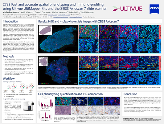
Fast and accurate spatial phenotyping and immuno-profiling using Ultivue UltiMapper kits and the ZEISS Axioscan 7 slide scanner
Register here for download
You are currently viewing a placeholder content from HubSpot. To access the actual content, click the button below. Please note that doing so will share data with third-party providers.
More InformationRelevant for
Collaboration, FixVUE, InSituPlex
Description Understanding the complexities of the tumor micro-environment in detail can vastly improve the accuracy of immuno-oncology research and accelerate the discovery of potential immunotherapy targets. Multiplexed immunofluorescence (mIF) assays have emerged as a critical approach for identifying complex cellular phenotypes in the tumor micro-environment, powered by the value of the spatial correlation of such phenotypes in a tissue specimen. Although many methods for multiplexed IF exist, they are often costly and require time-consuming assay development and long imaging times. The Ultivue UltiMapper kits enable rapid, pre-optimized staining of multiple targets simultaneously in a single FFPE tissue section. This technology is ready-to-use with conventional automated staining workflows and commercially available automated imaging systems. Here, we demonstrate the potential of accurate cellular phenotyping using the InSituPlex technology with fast whole-slide scanning using ZEISS Axioscan.Z1 an automated slide scanning system for fluorescence and transmitted light applications with modern cameras, a sophisticated focusing method and a powerful imaging software. The compounded effect of integrating these technologies can significantly improve the throughput of immuno-oncology research. A 4-target immunostaining assay was performed on multiple FFPE tumor sections using UltiMapper reagents. Briefly, slides were stained with a cocktail of primary antibodies using a Leica Biosystems BOND RX autostainer. Post-staining, the slides were coverslipped and imaged on ZEISS Axioscan.Z1. The resulting images were analyzed using IndicaLabs HALO analysis software. In this poster, we report on an efficient and streamlined workflow for mIF staining, imaging, and analysis of tumor samples. After nuclear segmentation of the resulting images, T-cell and macrophage populations were subtyped and identified through spatial identification of relevant biomarkers. The combined workflow presented here allows a user to get 4-plex, whole slide immunofluorescence images of FFPE tumor tissues within 6 hours. The automated whole slide scanning capability of ZEISS Axioscan.Z1 also allows a user to image up to 100 slides with minimal external input. The combination of optimized, fast staining UltiMapper reagents with ZEISS Axioscan.Z1 automated whole-slide fluorescence scanner enables rapid, deep immunoprofiling within the spatial context of the tumor, empowering translational and immuno-oncology research.
Authors
Catherine Benson, Keith Wharton, Gourab Chatterjee, Markus Neumann, Volker Döring and Maël Manesse

Want to meet with us? Check out our upcoming events.
Related Content
High throughput tissue phenotyping and imaging of the tumor immune microenvironment using novel FlexVUE™multiplexed immunofluorescence assays
Presented at AACR 2022
Introduction of a robust workflow for the whole-slide acquisition and co-registration of multiplex immunofluorescence tissue images for analysis of a 9-color, 8-marker immunophenotyping assay
RareCyte, Inc., Indica Labs & Ultivue
