Presented at: AACR 2022
Relevant for
Collaboration, FixVUE, Imaging mass cytometry, InSituPlex
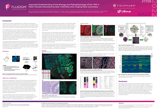
Description This poster demonstrates a unique tissue phenotyping workflow combining three complementary methods that can unravel novel insights in the complex tumor microenvironment. The highlighted workflow delivers tissue morphology information, spatial phenotyping of immune cell population on whole slides, and high-dimensional imaging in selected regions of interest (ROIs) by combining H&E, multiplex immunofluorescence (mIF), and Imaging Mass Cytometry™(IMC™) together with bespoke data analysis.
Andrew Quong, Nina Lane, Derek Quong, Gourab Chatterjee, Devan Fleury, Kirsteen H. Maclean, Maël Manesse, Keith A. Wharton Jr, Fabian Schneider, Dan Winkowski and James Mansfield.

Senior Product Manager AI & Data Analytics
Relevant for
FixVUE, InSituPlex
Description This poster demonstrates a unique tissue phenotyping workflow combining three complementary methods that can unravel novel insights in the complex tumor microenvironment. The highlighted workflow delivers tissue morphology information, spatial phenotyping of immune cell population on whole slides, and high-dimensional imaging in selected regions of interest (ROIs) by combining H&E, multiplex immunofluorescence (mIF), and Imaging Mass Cytometry™(IMC™) together with bespoke data analysis.
Moritz will drill his way through whatever intellectual challenge you’ll put in front of him. He previously perforated biochemistry, image analysis, data science, immuno-oncology biomarkers, product management/ownership. He’ll condense the best of our ideas into a coherent roadmap. His product backlog will guide us towards our vision one step at a time.
Senior Product Manager AI & Data Analytics
Relevant for
FlexVUE, InSituPlex
Description This poster demonstrates a unique tissue phenotyping workflow combining three complementary methods that can unravel novel insights in the complex tumor microenvironment. The highlighted workflow delivers tissue morphology information, spatial phenotyping of immune cell population on whole slides, and high-dimensional imaging in selected regions of interest (ROIs) by combining H&E, multiplex immunofluorescence (mIF), and Imaging Mass Cytometry™(IMC™) together with bespoke data analysis.
Moritz will drill his way through whatever intellectual challenge you’ll put in front of him. He previously perforated biochemistry, image analysis, data science, immuno-oncology biomarkers, product management/ownership. He’ll condense the best of our ideas into a coherent roadmap. His product backlog will guide us towards our vision one step at a time.
Moritz will drill his way through whatever intellectual challenge you’ll put in front of him. He previously perforated biochemistry, image analysis, data science, immuno-oncology biomarkers, product management/ownership. He’ll condense the best of our ideas into a coherent roadmap. His product backlog will guide us towards our vision one step at a time.
Senior Director Scientific Brand Strategy
Relevant for
FlexVUE, InSituPlex
Description This poster demonstrates a unique tissue phenotyping workflow combining three complementary methods that can unravel novel insights in the complex tumor microenvironment. The highlighted workflow delivers tissue morphology information, spatial phenotyping of immune cell population on whole slides, and high-dimensional imaging in selected regions of interest (ROIs) by combining H&E, multiplex immunofluorescence (mIF), and Imaging Mass Cytometry™(IMC™) together with bespoke data analysis.
Moritz will drill his way through whatever intellectual challenge you’ll put in front of him. He previously perforated biochemistry, image analysis, data science, immuno-oncology biomarkers, product management/ownership. He’ll condense the best of our ideas into a coherent roadmap. His product backlog will guide us towards our vision one step at a time.
Associate Director, Scientific Affairs Ultivue; Chief Science Officer, OracleBio
Relevant for
FixVUE, InSituPlex
Description This poster demonstrates a unique tissue phenotyping workflow combining three complementary methods that can unravel novel insights in the complex tumor microenvironment. The highlighted workflow delivers tissue morphology information, spatial phenotyping of immune cell population on whole slides, and high-dimensional imaging in selected regions of interest (ROIs) by combining H&E, multiplex immunofluorescence (mIF), and Imaging Mass Cytometry™(IMC™) together with bespoke data analysis.
Moritz will drill his way through whatever intellectual challenge you’ll put in front of him. He previously perforated biochemistry, image analysis, data science, immuno-oncology biomarkers, product management/ownership. He’ll condense the best of our ideas into a coherent roadmap. His product backlog will guide us towards our vision one step at a time.
Lorcan is an image analysis expert who previously spent 10 years in the pharmaceutical industry as a group leader (Organon, Schering-Plough, Merck & Co.). He led a group responsible for performing histology, immunohistochemistry / image analysis and he has considerable experience in developing translational biomarker strategies for projects across various therapeutic areas. He is a Prince 2® qualified project manager and has extensive experience in study management.
Senior Manager, FAS, North America
Relevant for
FlexVUE, InSituPlex
Description This poster demonstrates a unique tissue phenotyping workflow combining three complementary methods that can unravel novel insights in the complex tumor microenvironment. The highlighted workflow delivers tissue morphology information, spatial phenotyping of immune cell population on whole slides, and high-dimensional imaging in selected regions of interest (ROIs) by combining H&E, multiplex immunofluorescence (mIF), and Imaging Mass Cytometry™(IMC™) together with bespoke data analysis.
Moritz will drill his way through whatever intellectual challenge you’ll put in front of him. He previously perforated biochemistry, image analysis, data science, immuno-oncology biomarkers, product management/ownership. He’ll condense the best of our ideas into a coherent roadmap. His product backlog will guide us towards our vision one step at a time.
Principal Software Engineer
Relevant for
FixVUE, FlexVUE, InSituPlex, U-VUE
Description This poster demonstrates a unique tissue phenotyping workflow combining three complementary methods that can unravel novel insights in the complex tumor microenvironment. The highlighted workflow delivers tissue morphology information, spatial phenotyping of immune cell population on whole slides, and high-dimensional imaging in selected regions of interest (ROIs) by combining H&E, multiplex immunofluorescence (mIF), and Imaging Mass Cytometry™(IMC™) together with bespoke data analysis.
Moritz will drill his way through whatever intellectual challenge you’ll put in front of him. He previously perforated biochemistry, image analysis, data science, immuno-oncology biomarkers, product management/ownership. He’ll condense the best of our ideas into a coherent roadmap. His product backlog will guide us towards our vision one step at a time.
Associate Director Scientific Affairs
Relevant for
FlexVUE, InSituPlex
Description This poster demonstrates a unique tissue phenotyping workflow combining three complementary methods that can unravel novel insights in the complex tumor microenvironment. The highlighted workflow delivers tissue morphology information, spatial phenotyping of immune cell population on whole slides, and high-dimensional imaging in selected regions of interest (ROIs) by combining H&E, multiplex immunofluorescence (mIF), and Imaging Mass Cytometry™(IMC™) together with bespoke data analysis.
Moritz will drill his way through whatever intellectual challenge you’ll put in front of him. He previously perforated biochemistry, image analysis, data science, immuno-oncology biomarkers, product management/ownership. He’ll condense the best of our ideas into a coherent roadmap. His product backlog will guide us towards our vision one step at a time.
Senior Scientists, Product Development
Relevant for
FlexVUE, InSituPlex
Description This poster demonstrates a unique tissue phenotyping workflow combining three complementary methods that can unravel novel insights in the complex tumor microenvironment. The highlighted workflow delivers tissue morphology information, spatial phenotyping of immune cell population on whole slides, and high-dimensional imaging in selected regions of interest (ROIs) by combining H&E, multiplex immunofluorescence (mIF), and Imaging Mass Cytometry™(IMC™) together with bespoke data analysis.
Moritz will drill his way through whatever intellectual challenge you’ll put in front of him. He previously perforated biochemistry, image analysis, data science, immuno-oncology biomarkers, product management/ownership. He’ll condense the best of our ideas into a coherent roadmap. His product backlog will guide us towards our vision one step at a time.
Presented at: AACR 2022
Relevant for
Collaboration, FixVUE, Imaging mass cytometry, InSituPlex
Description This poster demonstrates a unique tissue phenotyping workflow combining three complementary methods that can unravel novel insights in the complex tumor microenvironment. The highlighted workflow delivers tissue morphology information, spatial phenotyping of immune cell population on whole slides, and high-dimensional imaging in selected regions of interest (ROIs) by combining H&E, multiplex immunofluorescence (mIF), and Imaging Mass Cytometry™(IMC™) together with bespoke data analysis.
Andrew Quong, Nina Lane, Derek Quong, Gourab Chatterjee, Devan Fleury, Kirsteen H. Maclean, Maël Manesse, Keith A. Wharton Jr, Fabian Schneider, Dan Winkowski and James Mansfield.
Presented at: AACR 2022
Relevant for
Collaboration, FixVUE, InSituPlex
Description This poster demonstrates a unique tissue phenotyping workflow combining three complementary methods that can unravel novel insights in the complex tumor microenvironment. The highlighted workflow delivers tissue morphology information, spatial phenotyping of immune cell population on whole slides, and high-dimensional imaging in selected regions of interest (ROIs) by combining H&E, multiplex immunofluorescence (mIF), and Imaging Mass Cytometry™(IMC™) together with bespoke data analysis.
Senior Field Applications Scientist, Ultivue
Relevant for
FixVUE, InSituPlex
Description This poster demonstrates a unique tissue phenotyping workflow combining three complementary methods that can unravel novel insights in the complex tumor microenvironment. The highlighted workflow delivers tissue morphology information, spatial phenotyping of immune cell population on whole slides, and high-dimensional imaging in selected regions of interest (ROIs) by combining H&E, multiplex immunofluorescence (mIF), and Imaging Mass Cytometry™(IMC™) together with bespoke data analysis.
Moritz will drill his way through whatever intellectual challenge you’ll put in front of him. He previously perforated biochemistry, image analysis, data science, immuno-oncology biomarkers, product management/ownership. He’ll condense the best of our ideas into a coherent roadmap. His product backlog will guide us towards our vision one step at a time.
Associate Director, Scientific Affairs; Senior Scientist, Visiopharm
Relevant for
Collaboration, FixVUE, InSituPlex
Description This poster demonstrates a unique tissue phenotyping workflow combining three complementary methods that can unravel novel insights in the complex tumor microenvironment. The highlighted workflow delivers tissue morphology information, spatial phenotyping of immune cell population on whole slides, and high-dimensional imaging in selected regions of interest (ROIs) by combining H&E, multiplex immunofluorescence (mIF), and Imaging Mass Cytometry™(IMC™) together with bespoke data analysis.
Moritz will drill his way through whatever intellectual challenge you’ll put in front of him. He previously perforated biochemistry, image analysis, data science, immuno-oncology biomarkers, product management/ownership. He’ll condense the best of our ideas into a coherent roadmap. His product backlog will guide us towards our vision one step at a time.
Dr. Fabian Schneider is part of Visiopharm’s R&D and Product Management team, responsible for phenotyping products as well as service projects for custom APP development. Fabian has over 10 years of international experience in cancer biology and immuno-oncology, working in academic research labs, clinical research teams and computational pathology groups in both academia and biopharma. Fabian received his Dr phil. nat. in Cell Biology in 2011 from the Johan Wolfgang Goethe University Frankfurt, Germany.

Relevant for
Collaboration, FixVUE, InSituPlex
Description We describe the potential of accurate cellular phenotyping using the InSituPlex technology with fast whole-slide scanning using ZEISS Axioscan 7, an automated slide scanning system for fluorescence and transmitted light applications with modern cameras, a sophisticated focusing method and a powerful imaging software. The compounded effect of integrating these technologies can significantly improve the throughput of immuno-oncology research.
Catherine Benson, Keith Wharton, Gourab Chatterjee, Markus Neumann, Volker Döring and Maël Manesse
Presented at: AACR 2021
Relevant for
Collaboration, FixVUE, InSituPlex
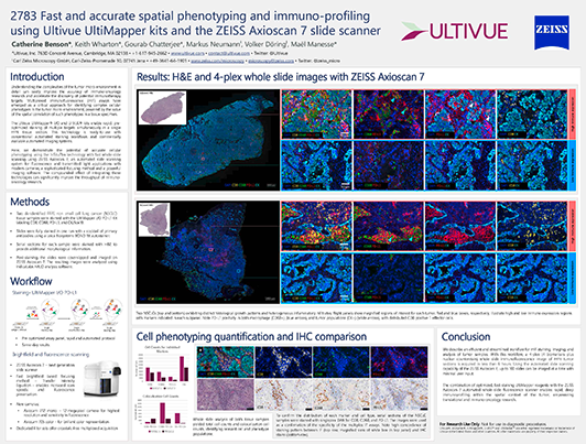
Description Understanding the complexities of the tumor micro-environment in detail can vastly improve the accuracy of immuno-oncology research and accelerate the discovery of potential immunotherapy targets. Multiplexed immunofluorescence (mIF) assays have emerged as a critical approach for identifying complex cellular phenotypes in the tumor micro-environment, powered by the value of the spatial correlation of such phenotypes in a tissue specimen. Although many methods for multiplexed IF exist, they are often costly and require time-consuming assay development and long imaging times. The Ultivue UltiMapper kits enable rapid, pre-optimized staining of multiple targets simultaneously in a single FFPE tissue section. This technology is ready-to-use with conventional automated staining workflows and commercially available automated imaging systems. Here, we demonstrate the potential of accurate cellular phenotyping using the InSituPlex technology with fast whole-slide scanning using ZEISS Axioscan.Z1 an automated slide scanning system for fluorescence and transmitted light applications with modern cameras, a sophisticated focusing method and a powerful imaging software. The compounded effect of integrating these technologies can significantly improve the throughput of immuno-oncology research. A 4-target immunostaining assay was performed on multiple FFPE tumor sections using UltiMapper reagents. Briefly, slides were stained with a cocktail of primary antibodies using a Leica Biosystems BOND RX autostainer. Post-staining, the slides were coverslipped and imaged on ZEISS Axioscan.Z1. The resulting images were analyzed using IndicaLabs HALO analysis software. In this poster, we report on an efficient and streamlined workflow for mIF staining, imaging, and analysis of tumor samples. After nuclear segmentation of the resulting images, T-cell and macrophage populations were subtyped and identified through spatial identification of relevant biomarkers. The combined workflow presented here allows a user to get 4-plex, whole slide immunofluorescence images of FFPE tumor tissues within 6 hours. The automated whole slide scanning capability of ZEISS Axioscan.Z1 also allows a user to image up to 100 slides with minimal external input. The combination of optimized, fast staining UltiMapper reagents with ZEISS Axioscan.Z1 automated whole-slide fluorescence scanner enables rapid, deep immunoprofiling within the spatial context of the tumor, empowering translational and immuno-oncology research.
Catherine Benson, Keith Wharton, Gourab Chatterjee, Markus Neumann, Volker Döring and Maël Manesse


 Image analysis
5829
Improved Understanding of the Biology and Pathophysiology of the TME in PDAC Samples Revealed by Both InSituPlex and Imaging Mass Cytometry
Image analysis
5829
Improved Understanding of the Biology and Pathophysiology of the TME in PDAC Samples Revealed by Both InSituPlex and Imaging Mass Cytometry
