

Presented at: AACR 2024
You are currently viewing a placeholder content from HubSpot. To access the actual content, click the button below. Please note that doing so will share data with third-party providers.
More InformationRelevant for
InSituPlex, mIF-H&E fusion
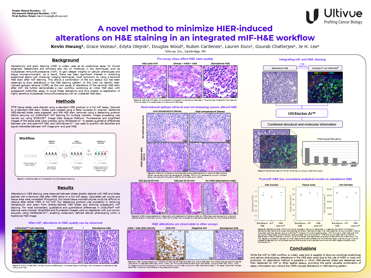
Description Discover a new workflow that enables you to effectively combine H&E staining with mIF InSituPlex® assays and avoid the heat-induced epitope retrieval (HIER) steps that are the root cause of altered H&E staining. We present data showing co-registration of H&E and mIF images with sub-micron accuracy, enabling highly accurate molecular characterization and cellular phenotyping.
Kevin Hwang, Grace Vezeau, Edyta Olejnik, Douglas Wood, Ruben Cardenes, Lauren Duro, Gourab Chatterjee, Je H. Lee

Presented at: AACR 2024
You are currently viewing a placeholder content from HubSpot. To access the actual content, click the button below. Please note that doing so will share data with third-party providers.
More InformationRelevant for
Spatial Image Analysis, Spatial Insights, STARVUE™, UltiAnalyzer.AI™
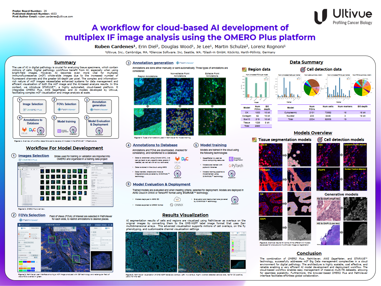
Description We present a highly scalable, cost-effective, and reliable AI workflow built with a combination of OMERO Plus, PathViewer, AWS SageMaker, and Ultivue software to efficiently manage mIF big data for digital pathology.
Ruben Cardenes, Erin Diel, Douglas Wood, Je Lee, Martin Schulze, Lorenz Rognoni
Presented at: AACR 2024
You are currently viewing a placeholder content from HubSpot. To access the actual content, click the button below. Please note that doing so will share data with third-party providers.
More InformationRelevant for
InSituPlex, OmniVUE™, U-VUE
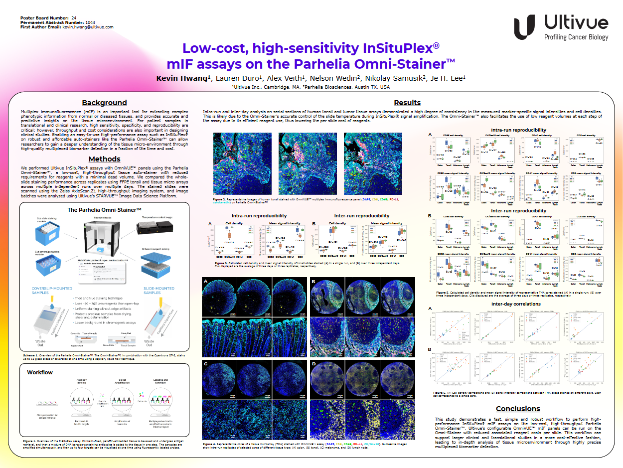
Description Our poster presents a simple, rapid, and robust workflow for performing Ultivue InSituPlex® mIF assays with OmniVUE™ panels on the Parhelia Omni-Stainer™. Tight slide temperature control and lower volume reagent used by the Omni-Stainer™ supports consistent, high performance, high throughput whole slide multiplexed biomarker detection for translational and clinical studies.
Kevin Hwang, Laura Duro, Alex Veith, Nelson Wedin, Nikolay Samisik, Je H. Lee.
Presented at: AACR 2024
You are currently viewing a placeholder content from HubSpot. To access the actual content, click the button below. Please note that doing so will share data with third-party providers.
More InformationRelevant for
Spatial Image Analysis, STARVUE™, UltiAnalyzer.AI™
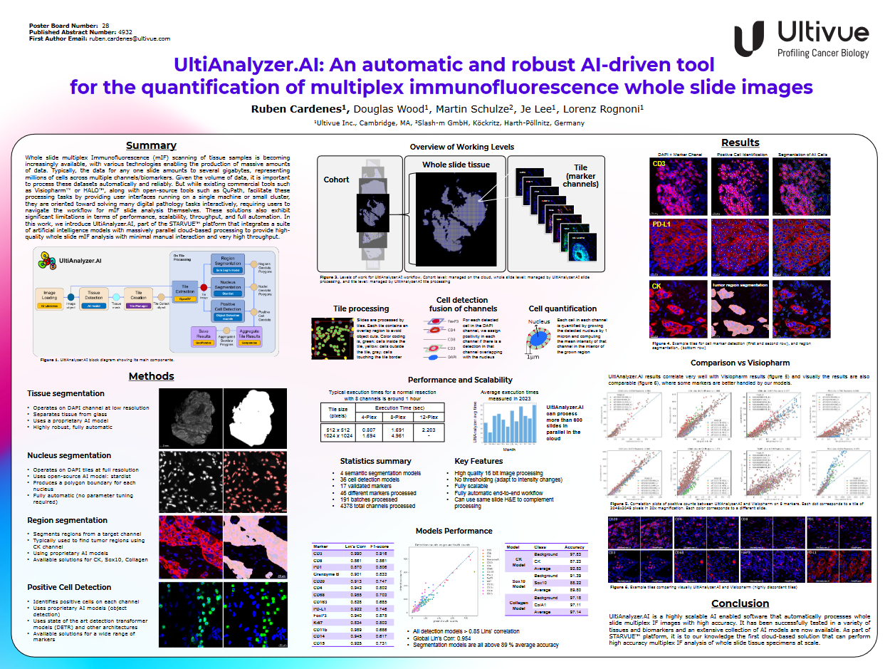
Description We introduce UltiAnalyzer.AI™, a software tool that integrates a set of artificial intelligence (AI) models designed for fully automatic, highly accurate, highly efficient, and fully scalable whole-slide mIF analysis. By utilizing the wide dynamic range of Ultivue’s InSituPlex® technology and cloud computing capabilities, this tool increases the robustness, reproducibility, and throughput of mIF analysis in digital pathology.
Ruben Cardenes, Douglas Wood, Martin Schulze, Je Lee, Lorenz Rognoni.

Director of Product Development at Ultivue, Inc. & Director of Scientific Affairs, Ultivue
Relevant for
InSituPlex
Description Though widely recognized that mIF assays have critical implications for clinical translation, validation of mIF staining that allows for flexibility in biomarker selection is a barrier for mIF integration into clinical practice. Join Yvette Cajigas and Angela Vasaturo, experts in assay development and biomarker strategy, and learn about a simple approach to evaluate sensitivity and reproducibility – without sacrificing the flexibility needed to answer biological questions required for evaluating specific treatment options. Take a deep dive into the world of multiplex immunofluorescence and see first-hand how Ultivue’s OmniVUE™ panels use its patented InSituPlex® technology to advance precision therapies in clinical trials.
Yvette Cajigas:
Yvette Cajigas is Director of Product Development at Ultivue, and is a board-certified histotechnologist with nearly 2 decades of experience in Anatomic Pathology. She has served as a technical lead at clinical academic and contract research pathology laboratories, and has extensive expertise in histologic technique assay development and multispectral digital pathology.
Angela Vasaturo, PhD
Dr. Angela Vasaturo is Director of Scientific Affairs at Ultivue. Prior to Ultivue, Angela was a Senior Researcher in Dr. Jerome Galon’s Laboratory of Integrative Cancer Immunology at the Cordeliers Research Center. Angela was among the first in Europe to be involved in the development of multiplex IHC and multispectral imaging and analysis of up to six immunofluorescence markers, and today is considered a leading European expert in multiplex IHC techniques, digital pathology, spatial biology, and tissue imaging.
![]()


Director of Scientific Affairs, Ultivue
Relevant for
InSituPlex
Description A persistent challenge in the field of immunotherapy is the fact that only a minority of patients respond to treatment. This is especially true for therapies which rely on immune activation, such as checkpoint inhibitors, due to the complex and heterogeneous immune escape mechanisms which can develop in each patient. Therefore, the development of robust biomarkers, which enable rational patient selection and the design of precise combination therapies, is key for the continued success and improvement of cancer treatments. Multiplex immunofluorescence (mIF) has emerged to complement the conventional IHC by providing highly reproducible, efficient, quantitative, and standardized assays for the analysis of formalin-fixed paraffin-embedded (FFPE) tissue samples. The simultaneous antibody-based detection of multiple markers on a single tissue section can address the low tissue availability from precious and rare donors, establishing mIF as an important assay in the study of the tumor microenvironment and one which can help to maximize the opportunity for patients to benefit from personalized immunotherapies. To enable the transition of mIF assays into clinical applications, a flexible, easy- to- use, and reproducible assay is needed to identify predictive biomarkers. Multiplex immunofluorescence gives researchers the opportunity to study the spatial relationships in the tumor microenvironment, providing access to a wealth of data and information about its immune contexture. The purpose of this presentation is to convey to scientists and pathologists the reasons why reproducible assays are essential for biomarker discovery during clinical trials, how multiplex mIF assays are a great tool for immune profiling the tumor microenvironment, and how spatial tissue analysis can help to make data-driven decisions.
Dr. Angela Vasaturo is Director of Scientific Affairs at Ultivue. Prior to Ultivue, Angela was a Senior Researcher in Dr. Jerome Galon’s Laboratory of Integrative Cancer Immunology at the Cordeliers Research Center. Angela was among the first in Europe to be involved in the development of multiplex IHC and multispectral imaging and analysis of up to six immunofluorescence markers, and today is considered a leading European expert in multiplex IHC techniques, digital pathology, spatial biology, and tissue imaging.
Senior Field Application Scientist, Ultivue
Relevant for
InSituPlex
Description Immunotherapy has transformed the treatment of metastatic and recurrent solid tumors. Advances in technology in the past few years have created unprecedented opportunities to identify biomarkers of disease processes, especially by using multi-omics technologies and datasets to derive valid and useful signatures of disease. Despite these advances, today only a minority of patients respond to immunotherapies. Prediction of response to therapies such as checkpoint inhibitors that rely on activation of endogenous immune responses has been shown to be especially difficult due to complex and heterogeneous immune escape mechanisms in each patient. Increasing evidence suggests that measurement of robust biomarkers through spatial analysis of the tissue will be key to enable rational patient selection for an improved clinical trial process and design precise combination therapies. The urgency to discover and implement new biomarkers lays bare the need to integrate a variety of advanced tools to probe the dynamic nature of events happening in the tumor microenvironment (TME). Recently, technology updates integrating the use of multiplex immunohistochemistry/immunofluorescence (mIHC/IF) provides much needed insight into cellular composition, cellular functions, and cell-cell interactions. Importantly, recent studies have used mIHC/IF to explore specific immune cells as part of the tumor immune microenvironment (TME) and found that it is helpful for clinical prognosis and efficacy prediction in patients with cancer. In this presentation we will show a streamlined unique workflow supporting whole slide imaging of an 8-plex mIF and traditional same slide H&E fusion on a single tissue slide for a comprehensive tissue immunophenotyping analysis.
Troy is an experienced analytical histotechnologist with expertise in histology, immunohistochemistry, in situ hybridization, digital slide scanning and image analysis within the GLP/GCP and pre-clinical environments along with a strong multidisciplinary CDx and research experience in models of oncology and autoimmune diseases.

Sr. Director Molecular & Computational Pathology, Ultivue
Relevant for
InSituPlex
Description Immunotherapy has transformed the treatment of metastatic and recurrent solid tumors but is challenged in that only a minority of patients respond, especially those therapies that rely on immune activation such as checkpoint inhibitors due to the complex and heterogeneous immune escape mechanisms which can develop in each patient. Therefore, the development of robust biomarkers, enabling rational patient selection and the design of precise combination therapies, is key for the continued success and improvement afforded by this valuable treatment. Overview of what is addressed: Why spatial relationships are important in tumor biology. Examine specific cell clustering, dispersion, and co-localization. To better understand the biology within tissues with a quantitative summary of the regions and phenotypes present.
As a Senior Director, Molecular and Computational Pathology at UV I am responsible for establishing augmented pathology workflows and for implementing innovative data-driven QC methods. I am a board-certified MD, PhD anatomical pathologist with 10 years of experience in Translational Research, and Cancer Diagnostics.
I am passionate about building cross-disciplinary bridges to deliver innovative services and products while considering the applicable regulatory requirements. I am excited about new ways that molecular and spatial biomarkers can accelerate pharmaceutical research and improve the lives of patients.

Associate Director Scientific Affairs, Ultivue; Chief Science Officer, OracleBio
Relevant for
InSituPlex
Description Immunotherapy has transformed the treatment of metastatic and recurrent solid tumors but is challenged in that only a minority of patients respond, especially those therapies that rely on immune activation such as checkpoint inhibitors due to the complex and heterogeneous immune escape mechanisms which can develop in each patient. Therefore, the development of robust biomarkers, enabling rational patient selection and the design of precise combination therapies, is key for the continued success and improvement afforded by this valuable treatment. Overview of what is addressed: Why spatial relationships are important in tumor biology. Examine specific cell clustering, dispersion, and co-localization. To better understand the biology within tissues with a quantitative summary of the regions and phenotypes present.
As a Senior Director, Molecular and Computational Pathology at UV I am responsible for establishing augmented pathology workflows and for implementing innovative data-driven QC methods. I am a board-certified MD, PhD anatomical pathologist with 10 years of experience in Translational Research, and Cancer Diagnostics.
I am passionate about building cross-disciplinary bridges to deliver innovative services and products while considering the applicable regulatory requirements. I am excited about new ways that molecular and spatial biomarkers can accelerate pharmaceutical research and improve the lives of patients.
Lorcan is an image analysis expert who previously spent 10 years in the pharmaceutical industry as a group leader (Organon, Schering-Plough, Merck & Co.). He led a group responsible for performing histology, immunohistochemistry / image analysis and he has considerable experience in developing translational biomarker strategies for projects across various therapeutic areas. He is a Prince 2® qualified project manager and has extensive experience in study management.


You are currently viewing a placeholder content from HubSpot. To access the actual content, click the button below. Please note that doing so will share data with third-party providers.
More InformationRelevant for
Collaboration, InSituPlex, OmniVUE (formerly FixVUE), OmniVUE (formerly FlexVUE)
Description Developing a multiplex immunofluorescence assay for the in depth characterization of activatedT cells in tumor tissue samples.
Director of R&D and Innovation, Ultivue
Relevant for
InSituPlex
Description Immunotherapy has transformed the treatment of metastatic and recurrent solid tumors but is challenged in that only a minority of patients respond, especially those therapies that rely on immune activation such as checkpoint inhibitors due to the complex and heterogeneous immune escape mechanisms which can develop in each patient. Therefore, the development of robust biomarkers, enabling rational patient selection and the design of precise combination therapies, is key for the continued success and improvement afforded by this valuable treatment. Overview of what is addressed: Why spatial relationships are important in tumor biology. Examine specific cell clustering, dispersion, and co-localization. To better understand the biology within tissues with a quantitative summary of the regions and phenotypes present.
As a Senior Director, Molecular and Computational Pathology at UV I am responsible for establishing augmented pathology workflows and for implementing innovative data-driven QC methods. I am a board-certified MD, PhD anatomical pathologist with 10 years of experience in Translational Research, and Cancer Diagnostics.
I am passionate about building cross-disciplinary bridges to deliver innovative services and products while considering the applicable regulatory requirements. I am excited about new ways that molecular and spatial biomarkers can accelerate pharmaceutical research and improve the lives of patients.
Vice President, Medical Director, Ultivue
Relevant for
InSituPlex
Description Immunotherapy has transformed the treatment of metastatic and recurrent solid tumors but is challenged in that only a minority of patients respond, especially those therapies that rely on immune activation such as checkpoint inhibitors due to the complex and heterogeneous immune escape mechanisms which can develop in each patient. Therefore, the development of robust biomarkers, enabling rational patient selection and the design of precise combination therapies, is key for the continued success and improvement afforded by this valuable treatment. Overview of what is addressed: Why spatial relationships are important in tumor biology. Examine specific cell clustering, dispersion, and co-localization. To better understand the biology within tissues with a quantitative summary of the regions and phenotypes present.
As a Senior Director, Molecular and Computational Pathology at UV I am responsible for establishing augmented pathology workflows and for implementing innovative data-driven QC methods. I am a board-certified MD, PhD anatomical pathologist with 10 years of experience in Translational Research, and Cancer Diagnostics.
I am passionate about building cross-disciplinary bridges to deliver innovative services and products while considering the applicable regulatory requirements. I am excited about new ways that molecular and spatial biomarkers can accelerate pharmaceutical research and improve the lives of patients.
Presented at: ASCO 2022
You are currently viewing a placeholder content from HubSpot. To access the actual content, click the button below. Please note that doing so will share data with third-party providers.
More InformationRelevant for
Collaboration, FixVUE, InSituPlex
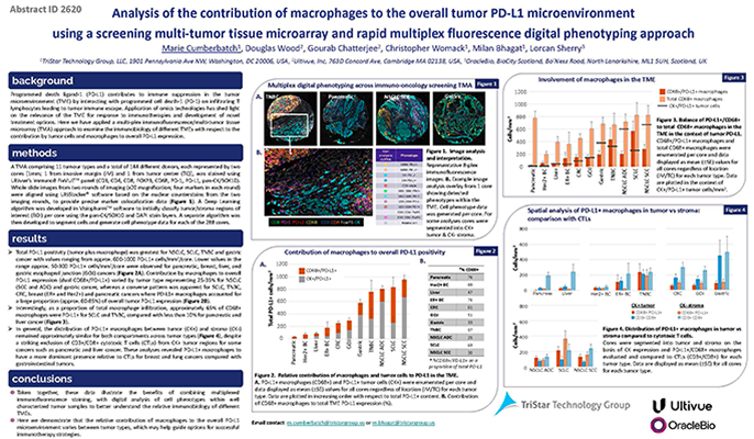
Description Marie Cumberbatch from TriStar Technology Group presented a poster of collaborative work between TriStar, Ultivue, and OracleBio at American Society of Clinical Oncology (ASCO) 2022 over the weekend. The work involved multiplex IF staining and analysis of multi-tumor tissue microarrays, with this poster focusing on the contribution of macrophages to the overall tumor PD-L1 microenvironment across different cancer types. Such target prevalence data can help guide options for successful immunotherapy strategies.
Marie Cumberbatch, Douglas Wood, Gourab Chatterjee, Christopher Womack, Milan Bhagat, Lorcan Sherry
Principal Pathologist-Scientist, Genentech
Relevant for
FixVUE, InSituPlex
Description Inhibitors of the programmed cell death-1 (PD-1/PD-L1) signaling axis are approved to treat non-small cell lung cancer (NSCLC) patients. However, most NSCLC patients do not respond to PD-(L)1 blockade as single agents, and intratumoral immune infiltrates involved in the response to these therapies remain poorly characterized. There remains a significant need to understand the biology of response and resistance and the role of infiltrating immune cells. Recent studies have suggested an association between increased B cell infiltration, along with the presence of tertiary lymphoid structures (TLSs) and improved response to immunotherapy in tumors from melanoma, soft tissue sarcoma, and renal cell carcinoma patients. Herein, the webinar will discuss whether intratumoral B cells are beneficial specifically in the context of PD-(L)1 blockade or are a general marker of a better prognosis in metastatic NSCLC.
Jena Giltnane is a translational pathologist-scientist in Genentech’s division of research and early development, where she leads digital and spatial pathology in support of translational oncology and cancer immunology programs through the use of multiplexed immunofluorescence tissue assays, tissue technology evaluation, and the collaborative development of deep learning based image analysis models in digital pathology.
Dr. Giltnane completed her MD, PhD, at Yale University in the laboratory of Dr. David Rimm and postdoctoral training in the laboratory of Dr. Carlos Arteaga at Vanderbilt University, where she also completed her Residency in Anatomic and Clinical Pathology.
She is an expert in genomic and proteomic biomarkers of diagnosis, prediction, and prognosis in breast cancer and the curation and analysis of high-dimensional clinical data.
You are currently viewing a placeholder content from Facebook. To access the actual content, click the button below. Please note that doing so will share data with third-party providers.
More InformationYou need to load content from reCAPTCHA to submit the form. Please note that doing so will share data with third-party providers.
More InformationYou are currently viewing a placeholder content from Hubspot Embedded Content. To access the actual content, click the button below. Please note that doing so will share data with third-party providers.
More InformationYou are currently viewing a placeholder content from HubSpot. To access the actual content, click the button below. Please note that doing so will share data with third-party providers.
More InformationYou are currently viewing a placeholder content from Hubspot Meetings. To access the actual content, click the button below. Please note that doing so will share data with third-party providers.
More InformationYou are currently viewing a placeholder content from Instagram. To access the actual content, click the button below. Please note that doing so will share data with third-party providers.
More InformationYou are currently viewing a placeholder content from X. To access the actual content, click the button below. Please note that doing so will share data with third-party providers.
More Information


 Spatial biology
10716
Combining Vizgen’s MERSCOPE® spatial transcriptomics and InSituPlex® spatial proteomics profiling to unravel the complexities of the tumor-immune microenvironment
Mary Bowen
Categories:
9248
A novel method to minimize HIER-induced alterations on H&E staining in an integrated mIF-H&E workflow
Spatial biology
10716
Combining Vizgen’s MERSCOPE® spatial transcriptomics and InSituPlex® spatial proteomics profiling to unravel the complexities of the tumor-immune microenvironment
Mary Bowen
Categories:
9248
A novel method to minimize HIER-induced alterations on H&E staining in an integrated mIF-H&E workflow
